
An adult human brain contains approximately 86 billion neurons, along with about 85 billion non-neuronal cells. All those cells work together to manage motor control, sensory perception, heart rate, breathing, and more. While the human brain is extremely complex, with the ability to process language, it is not totally unique. Most vertebrate brains are quite similar; they all need similar abilities, such as controlling body movements, controlling circadian rhythm, etc., to survive and reproduce. That means that there is the potential to learn about how the human brain works by studying other animals.
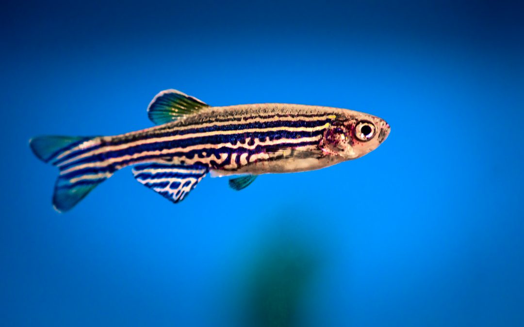
Scientists are using that approach by studying zebrafish to try to better understand how the brain develops. Scientists at Cornell University developed a technique for imaging the brain of an adult zebrafish, with the goal of better understanding human brain disorders. Zebrafish are easy to study when they are young, since their bodies are transparent, but that is not the case for adults. However, nerve cells in the brain produce calcium when activated, which gives scientists an angle to approach for imaging. The scientists at Cornell engineered zebrafish to produce a protein that binds to the calcium and fluoresces when exposed to a certain wavelength of light (480 nm). The fluorescent protein can then be imaged with a microscope, allowing the researchers to track when neurons are activated in adult zebrafish.
Previous applications of this technique had a problem, though. When the 480 nm laser passes through other fluorescent proteins, it excites them as well, which results in a blurry image. The new technique gets around this by using a longer wavelength beam (1400 nm), but focuses 3 beams on a single point in the brain. The wavelengths of the individual beams are too long to excite proteins by themselves, but when they combine at the focal point, they are strong enough to energize the target proteins. By running the lasers in a line across the brain, the researchers can stitch together a 2D image of brain activity. Running multiple of these cross sections allows the scientists to create 3D images of nerve activity in zebrafish brain structures.
There are several possible uses for this type of study. Researchers could genetically engineer fish to develop versions of human brain disorders (e.g. autism) and see how the brain is affected by the disease’s progression. Zebrafish could also be used as a model to study potential treatments for brain disorders. Scientists could follow the condition of the fish and see if brain condition and/or function improves during a proposed treatment.
Our refrigerated incubators are well suited for zebrafish studies like this one. They have a temperature range of 2-50°C and can easily maintain the warm zebrafish environment. They can also be adapted to create environments requiring vibration resistance, complete darkness, intense lighting, or a combination of variables. A microprocessor controls and displays the chamber temperature, and high and low temperature mechanical failsafes protect chamber contents in the unlikely event of a temperature excursion. For zebrafish applications, we use cool white (5000K color) LED lights. The light from these LEDs is most intense in the blue spectrum that is favorable for zebrafish rearing.
For more information on our refrigerated incubators, see our product page or contact us to request a catalog or quote.